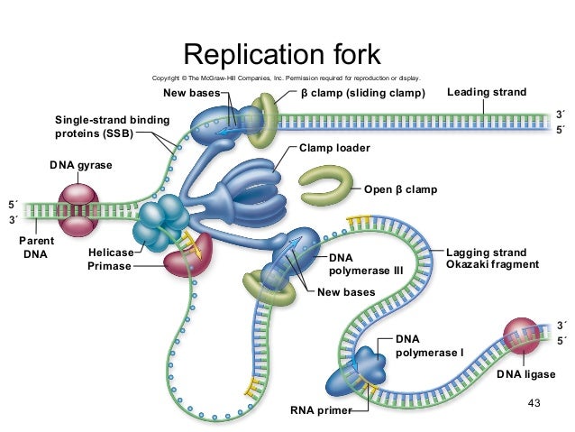
43 replication fork diagram labeled
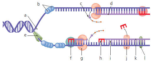
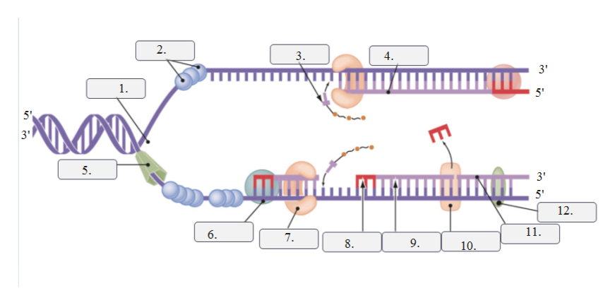
Correctly label the items in this DNA replication fork diagram. Drag the appropriate labels to their respective targets. Labels can be used more than once. Reset | Help Supercoiled chromosome Leading strand DNA polymerase III Lagging strand IIIIIIIIIIIIIIIIIIIIIIIIIIIII Pocong 5' Primase Copcodoro RNA primer Single-strand binding protein Helicase 3 Working with Molecular Genetics Chapter 5, DNA Replication I, v2 3 shifted to grow in media containing the highly abundant, light isotope of nitrogen, 14N, in the NH4Cl, so that newly synthesized DNA would have a "light" density.The labeled, heavy (old)
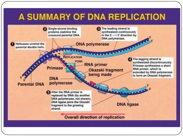
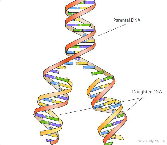
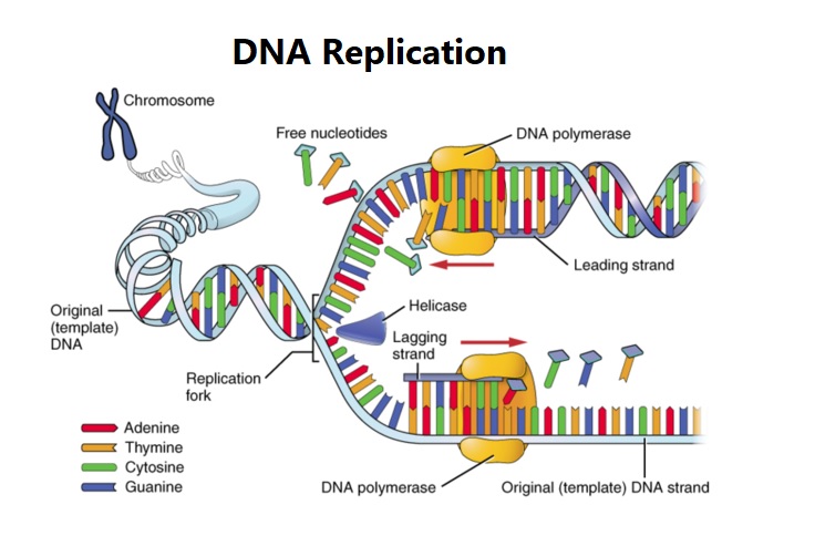
The detachment of the two single strands of DNA makes a two Y-formed design called a replication fork. Together they structure a bubble-like design called a replication bubble. These two separate strands fill in as a layout for the creation of the new DNA strands. Helicase is the main replication compound to be stacked at the beginning of ...

Replication fork diagram labeled
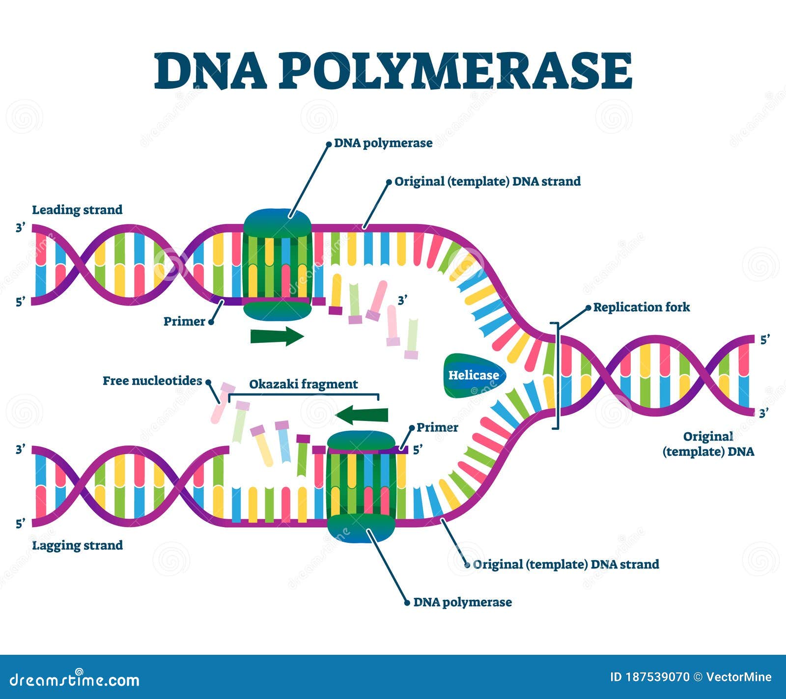
The elucidation of the structure of the double helix by James Watson and Francis Crick in 1953 provided a hint as to how DNA is copied during the process of DNA replication.Separating the strands of the double helix would provide two templates for the synthesis of new complementary strands, but exactly how new DNA molecules were constructed was still unclear. In this diagram of the process of DNA replication at a replication fork, the strand labeled B is the: DNA Replication The two antiparallel strands are replicated simultaneously in both directions. RNA primers are used to initiate a new strand. The parent strand at the 3' end of the template determines the Two replication forks are formed at the origin of replication, allowing for bidirectional replication and formation of a structure that looks like a bubble when viewed with a transmission electron microscope; as a result, this structure is called a replication bubble. The DNA near each replication fork is coated with single-stranded binding ...
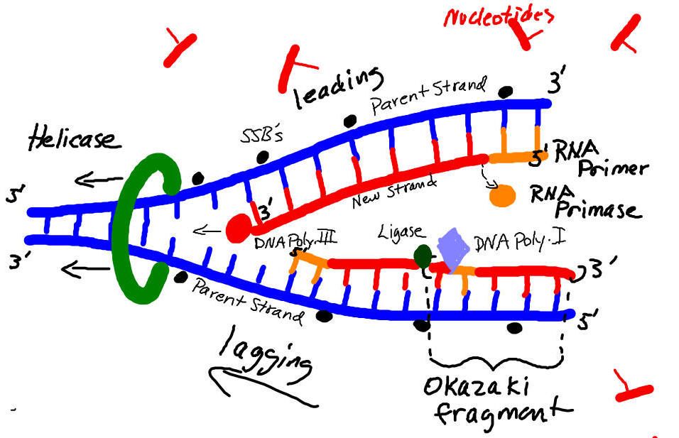
Replication fork diagram labeled. 1. On the diagram below, label the 5' and 3' ends of both parental DNA strands (you can make up which is which) 2. Label the replication fork 3. Draw and label helicase 4. Label the overall direction of DNA replication 5. Draw and label single stranded binding proteins 6. Draw and label the leading strand 7. corrects "overwinding" ahead of replication forks by breaking, swiveling, and rejoining DNA strands. Primase. An enzyme that creates a short RNA primer for initiation of DNA replication. RNA Primer. short segment of RNA used to initiate synthesis of a new strand of DNA during replication Label the diagram: DNA polymerase adds nucleotides (5' to 3') Replication fork is formed. DNA polymerase attaches to the primer. Okazaki fragments bound by ligase. DNA helicase unwinds DNA. Rearrange the steps to indicate the correct order: 1. Enzyme that unwinds DNA. Nov 12, 2018 · The diagram below shows a replication fork with the two parental DNA strands labeled at their 3' and 5 4/4(5). The diagram below shows a bacterial replication fork and its principal proteins. Drag the labels to their appropriate locations in the diagram to describe the name or function of each structure.
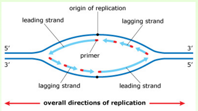
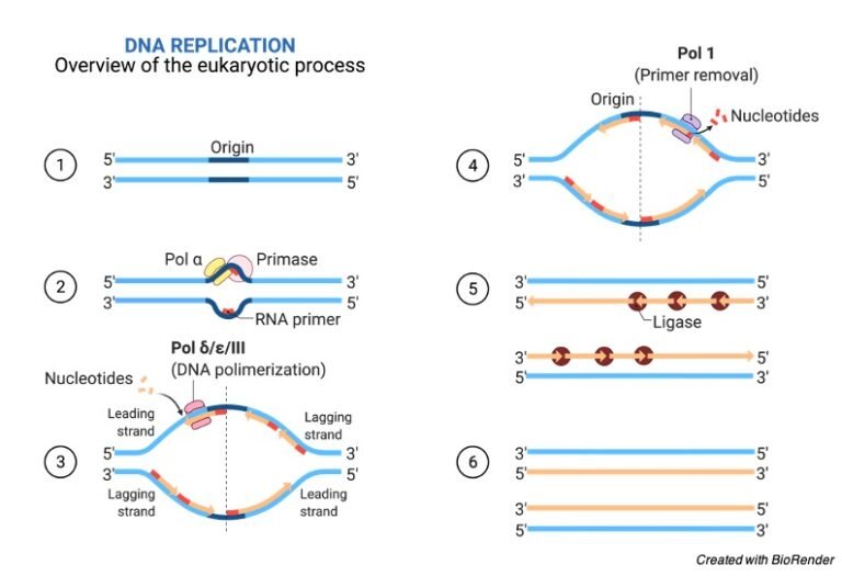
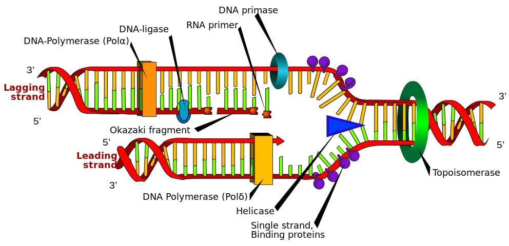
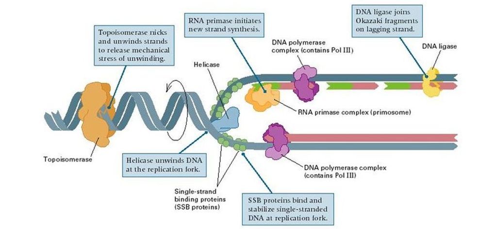
Replication Fork The replication fork * is a region where a cell's DNA * double helix has been unwound and separated to create an area where DNA polymerases and the other enzymes involved can use each strand as a template to synthesize a new double helix. DNA Base RNA Base An enzyme called a helicase * catalyzes strand separation. Label dna replication diagram. Leading and lagging strands okazaki fragment dna pol iii dna pol i dna ligase helicase primase single strand binding proteins rna primer replication fork and 5 and 3 ends of parental dna. Dna replication has three steps initiation elongation and termination. Multiple enzymes are used to complete this process ... As the DNA strands separate, a structure is created called the replication fork. The replication fork is the site at which DNA replication actually starts. Since DNA replication is bidirectional, that is it proceeds in both directions from the origin (Figure 3), there are actually two replication forks for each replication origin. Replication Forks The diagram below shows a bacterial replication fork and its principal proteins. Drag the labels to their appropriate locations in the diagram to describe the name or function of each structure. Use pink labels for the pink targets and blue labels for the blue targets. A) Breaks hydrogen bonds, unwinding DNA double helix.
Diagram of the Replication Fork. Brenda Grewe (public domain) Activity. ... The 5' and 3' ends of the parent DNA strands are labeled. The 3' end of a new strand has an arrow head. The fat arrow ... A moving replication fork. (A) This schematic diagram shows a current view of the arrangement of replication proteins at a replication fork when the fork is moving. The diagram in Figure 5-21 has been altered by folding the DNA on the lagging strand to ... replication fork: the Y-shaped structure formed during the initiation of replication semiconservative replication: the method used to replicate DNA in which the double-stranded molecule is separated and each strand acts as a template for a new strand to be synthesized, so the resulting DNA molecules are composed of one new strand of nucleotides ... Figure 1: Replication fork components. The RF is a multiprotein complex with helicase and DNA synthesis activities. It is called a fork because the structure resembles a two-pronged fork. The ...

The replication fork | USMLE Step 1 Forum
DNA replication is the process of duplication of DNA during cell division. DNA is a self-replicating structure and the semi-conservative process of DNA replication occurs in the sequence of initiation, elongation, and termination. The "unzipping" of the DNA double helix occurs by helicase enzyme which breaks the hydrogen bonds holding the ...

Chapter 9: DNA Replication - Chemistry
ColE1 Replication Control-an example of primer control of replication 1. RNAII will serve as a primer for the replication fork. 2. The 3’ end is processed by host RnaseH to allow efficient RNA-DNA hybrid to form 3. The hybrid acts as a primer for host Pol1 4. As the concentration of plasmid increases, Rop does also 5. Rop stabilizes the RNA1 ...
Dna replication
As the DNA opens, two Y-shaped structures called replication forks are formed, together making up what's called a replication bubble. The replication forks will move in opposite directions as replication proceeds. Diagram based on similar illustration in Reece et al. . How does replication actually get going at the forks?

Similiar Replication Fork Diagram Keywords | Dna molecule ...
The replication fork is a very active area where DNA replication takes place. It is created when DNA helicase unwinds the double helix structure of the DNA. The replication fork looks like a fork ...
What is a replication fork? - Quora
Problem 5: More on the replication fork In this diagram of the process of DNA replication at a replication fork, the black boxes labeled D and E are: A RNA primers. B DNA template strands. C Okazaki fragments. D DNA polymerase. E Newly synthesized DNA strand. The Biology Project

Pines
Jul 18, 2021 · Replication Fork. The replication fork is a structure which is formed during the long helical DNA during the process of DNA replication. It is activated by helicases, which helps in breaking the hydrogen bonds, which holds the two strands of the helix. The resulting structure has two branching’s which is known as prongs, where each one is ...

Solved: The Image Below Shows A Replication Fork With Temp ...
On the DNA diagram above, label the 5' and 3' ends of the template DNA strands by filling in the shaded boxes and show the direction of movement of the replication fork by an arrow. d) Label the parental strands as the templates for . leading (continuous) or . lagging (discontinuous) strand synthesis. e)

Media Portfolio
1. Below is a replication fork (one side of an origin): a double-stranded DNA partially opened up to provide single-stranded regions where replication can occur. Draw and label the following: RNA primers (label 3′ and 5′ ends), leading-strand DNA polymerase and new DNA (label 3′ and 5′ ends and show direction of synthesis
34 In The Replication Fork Below Label The Leading And ...
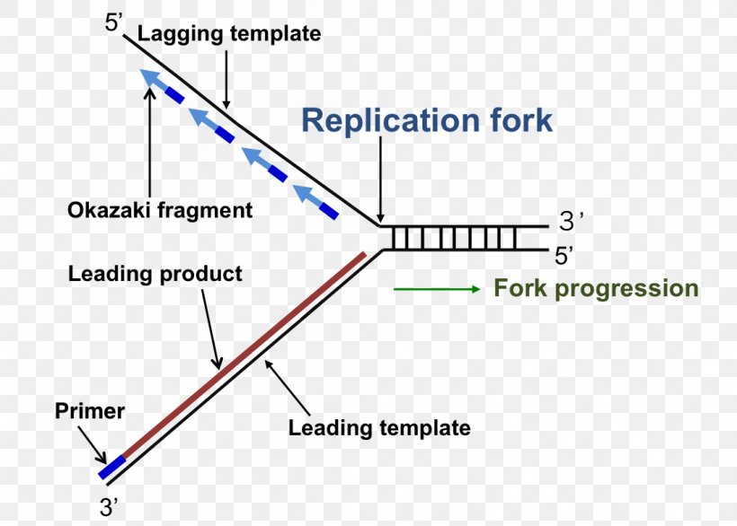
The replication fork. Leading-strand synthesis proceeds continuously in the 5' to 3' direction. Lagging-strand synthesis also occurs in the 5' to 3' direction, but in a discontinuous manner. An RNA/DNA primer (labeled in green) initiates leading-strand synthesis and every Okazaki fragment on the lagging strand.
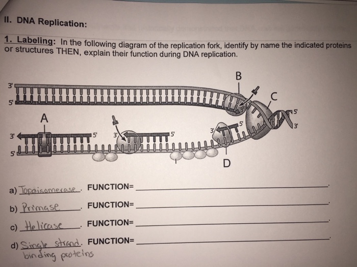
Solved: In The Following Diagram Of The Replication Fork ...
The advancing replication forks meet at a point opposite to the point of origin thus opening up the coiled DNA molecule. This is called θ (theta) model of replication. But the unwinding is a very complex mechanism as the two strands are coiled. The two advancing replication forks make the remaining entire un-replicated portion of DNA overwound.
Print Exam 3: Chs. 5 (DNA Structure and Replication ...
The strand, which is synthesized in the same direction as the replication fork, is known as the 'leading' strand. The template for this strand runs in the direction of 3' to 5'. The Polymerase has to attach only once and it can continue its work as the replication fork moves forward.
The process of DNA replication is illustarted in the ...
Two replication forks are formed at the origin of replication, allowing for bidirectional replication and formation of a structure that looks like a bubble when viewed with a transmission electron microscope; as a result, this structure is called a replication bubble. The DNA near each replication fork is coated with single-stranded binding ...

An Autumn Welcoming
In this diagram of the process of DNA replication at a replication fork, the strand labeled B is the: DNA Replication The two antiparallel strands are replicated simultaneously in both directions. RNA primers are used to initiate a new strand. The parent strand at the 3' end of the template determines the
Answered: Label the figure to assess your… | bartleby
The elucidation of the structure of the double helix by James Watson and Francis Crick in 1953 provided a hint as to how DNA is copied during the process of DNA replication.Separating the strands of the double helix would provide two templates for the synthesis of new complementary strands, but exactly how new DNA molecules were constructed was still unclear.
Replication Fork: Definition, Structure, Diagram, & Function
DNA Replication Replication Fork Enzyme Triangle, PNG ...
DNA Replication Process, Steps, Diagram, Enzymes ...

Why does dna replicate
Biology 201 Chapter 14

DNA Replication — Steps & Diagram - Expii

Enzymes and reactions in the DNA replication fork. Major ...

30 The Diagram Below Shows A Bacterial Replication Fork ...

Helicase-DNA polymerase at the replication fork | Download ...
DNA Polymerase Enzyme Syntheses Labeled Educational Vector ...
DNA Replication: Steps, Process, Diagram and Simple ...

Diagram Dna Replication Fork Labeled - Aflam-Neeeak

autumn mountain view

Solved: This Diagram Depicts An Origin Of Replication, Wit ...
Topic 7.1 DNA Structure and Replication - AMAZING WORLD OF ...

Chapter 16: DNA Structure and Replication
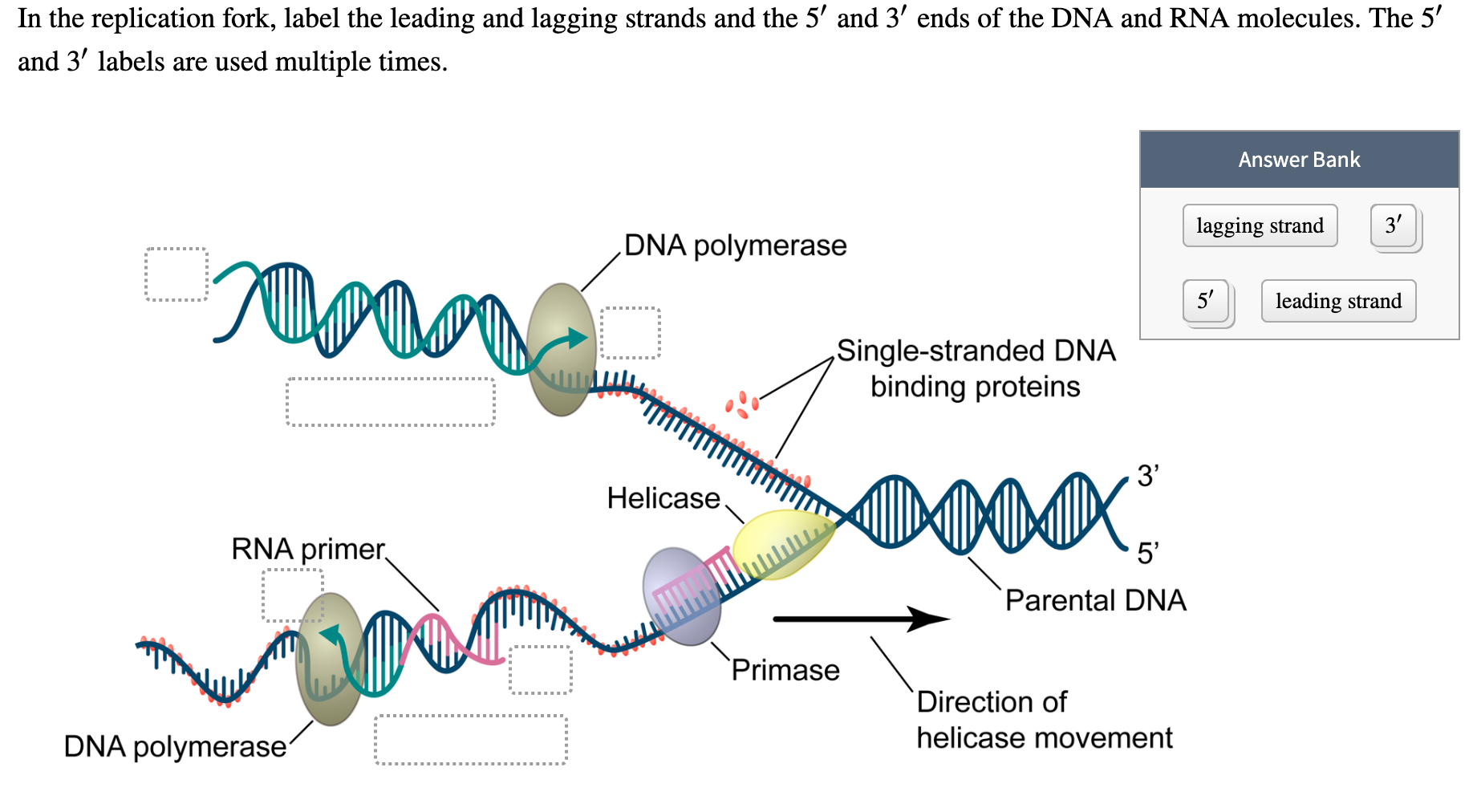
Solved: In The Replication Fork, Label The Leading And Lag ...

DNA replication | Molecular Biology | Pinterest | Genetics ...
Dna Replication Drawing at PaintingValley.com | Explore ...
small waterfall in a stream

Quia - 9AP Molecular Genetics Unit Basic Flashcards (ch 16-21)

The Diagram Below Shows A Bacterial Replication Fork And ...
Interaction of RecG with stalled replication fork ...

Origins of replication. A: A schematic drawing of a ...

DNA Replication ~ DNA & The Life

Model of a replication fork showing leading and lagging ...

Unlike the replication fork... | Download Scientific Diagram

DNA Replication Fork: Definition & Overview - Video ...
Replication fork re-start. | Download Scientific Diagram


Comments
Post a Comment