42 compound microscope ray diagram
October 15, 2019 - Click here👆to get an answer to your question ✍️ With the help of ray diagram, describe the construction, working of a compound microscope when the final image is formed at least distance of distinct vision (D = 25 cm) . Derive an expression fords magnifying power (m). (a) Draw a labelled ray diagram of compound microscope, when final image forms at the least distance of distinct vision. (b) Why is its objective of short focal length and of short aperture, compared to its eyepiece? Explain. (c) The focal length of the objective is 4 cm while that of eyepiece ...
1,837. 181. Summary: Compound microscope uses two convex lenses, one as objective lens and one as eye piece lens. What is the length of compound microscope? If we say "length of microscope", which distance does it refer to? Is it: a) the tube length (L), which is the distance between the focal point of objective and focal point of eye piece ...

Compound microscope ray diagram
Compound Microscope Ray Diagram Mistakes Physics Forums. Share this post. 1 Response to "Compound Microscope Diagram With Labels" Wayen June 15, 2021 at 4:16 AM. Excellent point, comparable writings are I don't have the foggiest idea whether they are in the same class as your work out. Microscopes were first developed in the early 1600s by eyeglass makers in The Netherlands and Denmark. The simplest compound microscope is constructed from two convex lenses as shown schematically in Figure 2. The first lens is called the objective lens, and has typical magnification values ... Simple Microscope Definition. A simple microscope is one that uses a single lens for magnification, such as a magnifying glass while a compound microscope uses several lenses to enhance the magnification of an object. It uses a lens to enlarge an object through angular magnification alone, giving the viewer an erect enlarged virtual image.
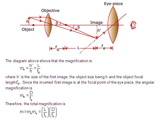
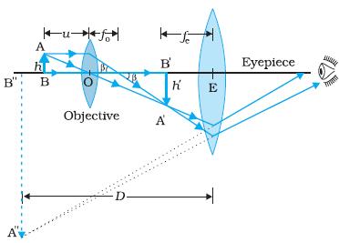
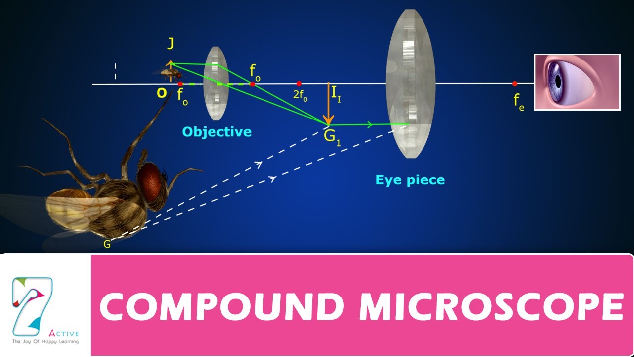
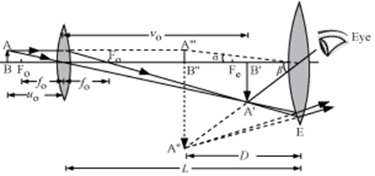
Compound microscope ray diagram. In the compound microscope, total magnification is the ocular lens magnification multiplied by the objective lens magnification. ... Draw ray diagram and apply thin lens formula. A) A compound ... Blank microscope diagram pdf wiring diagram for party switch u2022 rh lomond tw Microscope Diagram Worksheet Compound Microscope. There are two adjustment knobs, or flowers. The microscope compound light microscope parts of her husband in its own function properly in correctly now will cover information on. Draw a neat ray diagram to show the image formation in a compound microscope. Darfine its magnifying pieer and derive an expression for it when the final image is at near point of eye. asked Dec 22, 2021 in Physics by kiran5870 ( 20 points) A tiny object AB to be magnified is placed in front of the objective lens just beyond its principal focus fo’. In this case, the objective lens O of the compound microscope forms a real, inverted and enlarged image A’B’ of the object.
Sep 17, 2021 - ADVERTISEMENTS: Read this article to learn about the working principle and parts of a compound microscope with diagrams! Working Principle: The most commonly used microscope for general purposes is the standard compound microscope. It magnifies the size of the object by a complex ... (a) Labelled diagram of compound microscope. The objective lens form image A' B' near the first focal point of eyepiece. (b) Angular magnification of objective lens m 0 = linear magnification h'/h. where L is the distance between second focal point of the objective and first focal point of eyepiece.If the final image A'' B'' is formed at the near point. Ray Diagram Of A Compound Microscope Diagram Principles Complex Systems . Ray Optics And Optical Instruments Videos Refraction Internal Reflection Physics Notes Physics Projects Physics Classroom . Research The Topic What Are Concave And Convex Lenses Part 1 Light Science Physical Science Physics Classroom . Click here👆to get an answer to your question ✍️ Draw a ray diagram of compound microscope, when final image is formed at the minimum distance of distinct vision.
Dec 7, 2020 — draw a labelled ray diagram showing imagr formation in compound microscope.define its magnifying power and write expression for it. If you are not redirected automatically, follow the link to Funsciecne.in Deduce an expression for the total magnification when the final image is formed at the near point. In a compound microscope, an object is placed at a distance ... Draw a ray diagram of a compound microscope for the final image formed at least distance of distinct vision.
ABU-SARIM English blog: Ray Diagram for Compound microscope
Brightfield Microscope Definition. Brightfield Microscope is also known as the Compound Light Microscope. It is an optical microscope that uses light rays to produce a dark image against a bright background. It is the standard microscope that is used in Biology, Cellular Biology, and Microbiological Laboratory studies.

optics - Ray diagram of focussing on a compound microscope ...
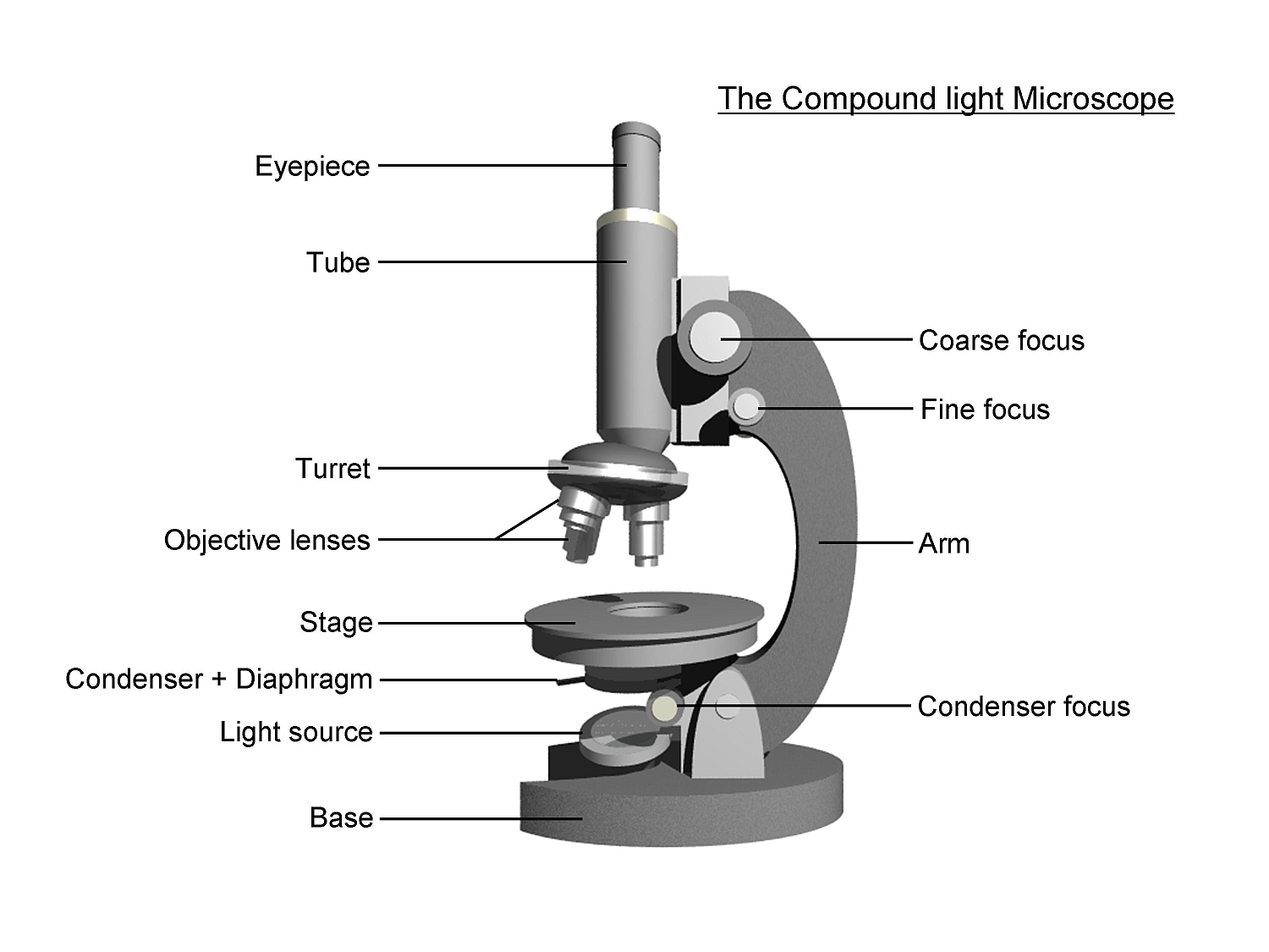
Compound microscope is a type of optical microscope that is used for obtaining a high-resolution image. There are more than two lenses in a compound microscope. Learn about the working principle, parts and uses of a compound microscope along with a labeled diagram here.
Labeled Microscope Lens Diagram - Micropedia
Do you need help with your homework? On Toppr Answr you can scan any question, and get its answer instantly

Metallurgical Microscopes - MicroscopeGenius.com
Draw a ray diagram to show the working of a compound microscope. Deduce an expression for the total magnification the final image is formed at the near point.
In a compound microscope , an object is placed at a distance of 1.5 cm from the objective of focal length 1.25cm. if the eye piece has a focal length of 5 cm and the final image is formed at the near point, estimate the magnifying ...

ICSE Class 8 Physics - Light - Compound Microscope Ray ...
(i) Draw a ray diagram showing the image formation by a compound microscope. Obtain expression for total magnification when the image is formed at infinity. (ii) How does the resolving power of a compound microscope get affected, when (a) Focal length of the objective is decreased. (b) The wavelength of light is increased?
a draw a ray diagram showing the image formation by a ...
Draw a ray diagram of compound microscope, when the final image is formed at the minimum distance of distinct vision. Hint: A compound microscope is an optical instrument used for observing highly magnified images of tiny objects. The compound microscope has two lenses and it works in such a way that the image of the object from the first lens ...
a draw a ray diagram to show the working of a compound ...
(a) Draw a ray diagram for final image formed at distance of distinct vision (D) by a compound microscope and write expression for its magnifying power. (b)An angular magnification (magnifying power) of 30x is desired for a compound microscope using as objective of focal length 1.25cm and eye piece of focal length 5cm.

Draw a ray diagram to show formation of an image by a ...
Advertisement for Students Studying Abroad in Class X of National Talent Search Examination - 2020 · Results of Selection Test of the Diploma Course in Guidance and Counselling (2022 session) · Observance of 'Armed Forces Flag Day' on 7.12.2021 & Contribution towards Armed Forces Flag Day Fund

(a) Draw the ray diagram showing the formation of toppr.com
(a) Draw a ray diagram for the formation of image by a compound microscope. (b) You are given the following three lenses. Which two lenses will you use as an eyepiece and as an objective to construct a compound microscope? Lenses Power (D) Aperture (cm) L1 3 8 L2 6 1 L3 10 1 (c) Define resolving power of a microscope and write one factor on which it depends.

Image from page 105 of "The Bell System technical journal" (1922)
September 16, 2016 - ADVERTISEMENTS: In this article we will discuss about:- 1. Essential Parts of Compound Microscope 2. Magnification of the Image of the Object by Compound Microscope 3. Resolution Power 4. Method for Studying Microbes 5. Measurement of the Size of Objects. Essential Parts of Compound Microscope: ...
black framed eyeglasses on white surface
Ray diagram of a compound microscope.When the final image is formed at the least distance of distinct vision,For the image formed at infinity, ue = feand By making focal length of the objective small, the magnifying power can be increased.

(a) Draw a labelled ray diagram of compound microscope ...
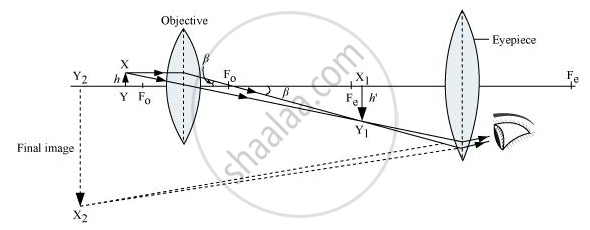
Ray diagram of Compound microscope. The specimen AB is placed just beyond the principal focus Fo' of the objective lens. A ray of light AO from A goes parallel to the principal axis towards the objective lens and converges.

Draw a ray diagram of a compound microscope for the final ...
May 19, 2017 · Here is the ray diagram of a compound microscope. So, when we are focussing, we move the objective lens which tweaks the image distance. My doubt is that, shouldn't the image be seen clearly, wheresoever the first real image forms, if within Fe (Focus of the eyepiece lens).

Image from page 369 of "Electron microscopy; proceedings of the Stockholm Conference, September, 1956" (1957)
October 15, 2019 - Click here👆to get an answer to your question ✍️ Describe the construction of a compound microscope. Derive an expression for its total magnification. Draw a ray diagram for the formation of image by a compound microscope.
Microscope Total Magnification Formula - Micropedia
This Fastest drawing Technique to draw Compound Microscope Ray Diagram is for all students of every Board - WBCHSE, HSC, CBSC, ICSE and other state boards fr...
Compound Microscope, Ray Diagram Mistakes. | Physics Forums
May 08, 2020 · A Draw A Ray Diagram Showing The Image Formation By A Compound. Magnification Of Compound Microscope With Derivation And Diagram. Solved The Focal Length Of A Compound Microscope S Object. Notes On Microscope Grade 11 Physics Optical Instruments. The Compound Microscope Physics Homework Help Physics Assignments.

a draw a ray diagram showing the image formation by a ...
(i) Draw a labelled ray diagram showing the formation of a final image by a compound microscope at the least distance of distinct vision. (ii) The total magnification produced by a compound microscope is 20. The magnification produced by the eye piece is 5. The microscope is focused on a certain object.

Image from page 155 of "The microscope; an introduction to microscopic methods and to histology" (1917)
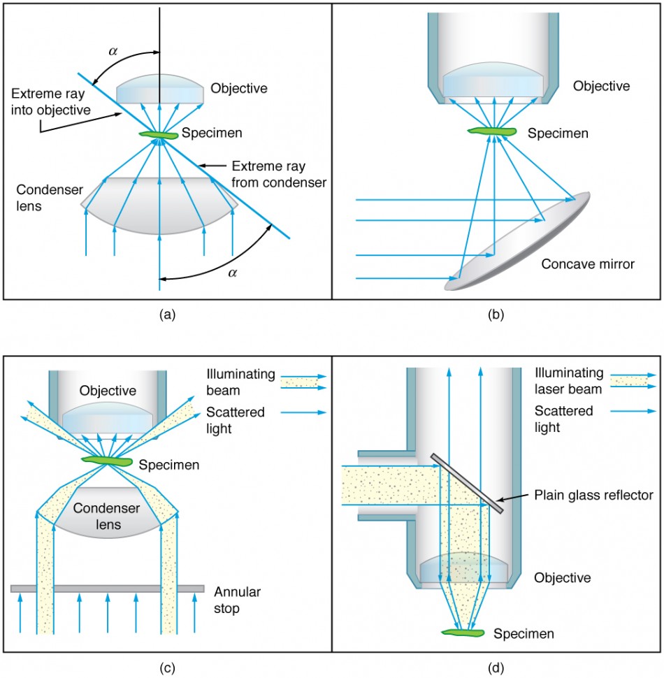
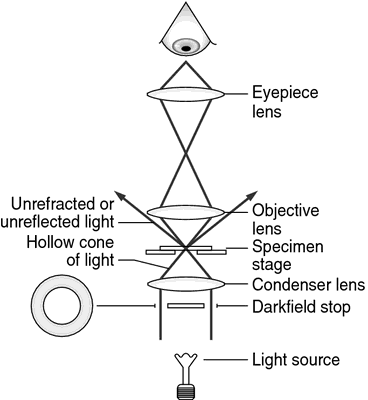
This is to say: The dark-ground microscopy makes use of the dark-ground microscope, a special type of compound light microscope. The dark-field condenser with a central circular stop, which illuminates the object with a cone of light, is the most essential part of the dark-ground microscope.

Compound Microscope Derivation Class 11 - Micropedia
icse class 8 physics light compound microscope ray. Microscope Ray Diagram. Here are a number of highest rated Microscope Ray Diagram pictures on internet. We identified it from reliable source. Its submitted by government in the best field. We put up with this kind of Microscope Ray Diagram graphic could possibly be the most trending topic ...
Compound Microscope Diagram Class 11 - Micropedia
(ii) A compound microscope has an objective of focal length 1.25 cm and eyepiece of focal length 5 cm. a small object is kept as 2.5 cm from the objective. If the final image formed is at infinity, find the distance between the objective and the eyepiece. ... Draw a labeled ray diagram to show the formation of image in an astronomical telescope ...

The compound microscope - how to draw ray diagrams - YouTube
The optical microscope often referred to as the light microscope, is a type of microscope that uses visible light and a system of lenses to magnify images of small subjects. There are two basic types of optical microscopes: Simple microscopes. Compound microscopes. The term "compound" in compound microscopes refers to the microscope having ...

brown concrete building during daytime
I am trying to draw the ray diagram of a compound microscope. Here is my attempt (sorry if the image is kind of blurry): I believe that I understand how the objective lens creates a real image (I

Image from page 299 of "A treatise on physiology and hygiene for educational institutions and general readers .." (1887)
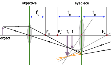
distance or image size. Thus, a ray diagram is used to find the final image distance and to determine the total magnification. The overall magnification of a compound microscope is the product of the individual magnifications of each lens: m = mome (1) where the magnification of either lens is given by: m = - q/p (2)

Compound Microscope Drawing - ClipArt Best
Ray Diagram of a Compound Microscope Complex Systems, Life Science, Biology, ... Ray Diagram for a Convex Lens showing the Focal Point and Focal Length.

white concrete building with blue windows
Here are two ray diagrams for compound microscope, the first one proposed by the book, and the second one recommended by the teacher: View attachment 232683 View attachment 232684 In the first image, the light rays form a real image A'B', which becomes the virtual object for the eyepiece. See, the original rays are carried forward to the ...

Important Questions for CBSE Class 12 Physics Optical ...
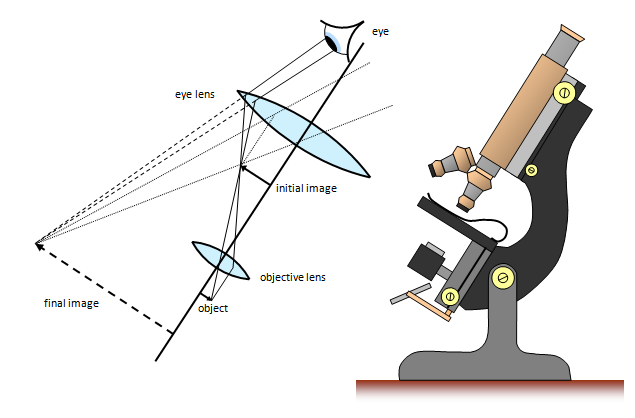
A compound microscope consists of two convex lenses coaxially separated by some distance. The lens nearer to the object is called the objective. The lens through which the final image is viewed is called the eyepiece. The focal length of objective lens is smaller than eyepiece. Ques. Draw a ray diagram of a compound microscope.

person swimming on top of Manta Ray fish
Click here to get an answer to your question ✍️ Draw the ray diagram of image formation in case of compound microscope.1 answer · Top answer: Ray diagram of image formation by a compound microscope is shown above.

Compound Microscope Diagram Class 12 Physics - Micropedia
Ray diagram of a compound microscope.When the final image is formed at the least distance of distinct vision,For the image formed at infinity, ue = feand By making focal length of the objective small, the magnifying power can be increased.
Draw a ray diagram for the formation of image by a ...
Here are a number of highest rated Simple Compound Microscope Diagram pictures upon internet. We identified it from well-behaved source. Its submitted by admin in the best field. We acknowledge this nice of Simple Compound Microscope Diagram graphic could possibly be the most trending topic later than we portion it in google lead or facebook.

Draw a labelled ray diagram to show image formation by a ...
eLearn · is the official repository of digitized textbooks. Each book has been augmented with Video Lectures, Illustrations, Animations, Simulations and Interactive Assessments. Through this website, you can access 30 Science and Maths textbooks for Grade 1-12 which have been augmented with ...
Science - Cellphone Microscope
Simple Microscope Definition. A simple microscope is one that uses a single lens for magnification, such as a magnifying glass while a compound microscope uses several lenses to enhance the magnification of an object. It uses a lens to enlarge an object through angular magnification alone, giving the viewer an erect enlarged virtual image.
(a) Draw a ray diagram showing the image formation by a ...
Microscopes were first developed in the early 1600s by eyeglass makers in The Netherlands and Denmark. The simplest compound microscope is constructed from two convex lenses as shown schematically in Figure 2. The first lens is called the objective lens, and has typical magnification values ...

Solved: Compound Microscope 2 1 2 3 4 In The Following Ray ...
Compound Microscope Ray Diagram Mistakes Physics Forums. Share this post. 1 Response to "Compound Microscope Diagram With Labels" Wayen June 15, 2021 at 4:16 AM. Excellent point, comparable writings are I don't have the foggiest idea whether they are in the same class as your work out.

grayscale photography of hospital
Compound microscope | definition of compound microscope by ...

gray concrete statue of a man
Drawing Compound Microscope Diagram With Labels - Micropedia

Simple Microscope Ray Diagram - Micropedia
Microscopes, Geometrical Optics - from A-level Physics Tutor

draw ray diagrams to show the image formation in a ...
X-ray microscope | definition of x-ray microscope by ...

Image from page 17 of "The microscope; an introduction to microscopic methods and to histology" (1901)


Comments
Post a Comment